Thyroid Nodules Explained: Benign vs Cancerous and Biopsy Guidelines
Understanding Your Thyroid Health
Imagine getting a call from your doctor saying you have a "lump" on your neck. Your heart races. You wonder immediately if it is cancer. Here is the truth that many people miss: thyroid nodules are incredibly common. In fact, if we used high-sensitivity ultrasound on everyone over 60, nearly two-thirds would have one. Most of these small lumps turn out to be completely harmless. However, about 5 to 10 percent do carry signs of thyroid cancer. The goal isn't just to find the problem, but to figure out exactly which ones matter.
The Anatomy of a Thyroid Nodule
Thyroid NoduleA thyroid nodule is a solid or fluid-filled lump that forms within the thyroid gland.
You likely feel nothing. Thyroid nodules are often discovered during a routine physical exam for something else, or sometimes found incidentally on a CT scan of your neck for dental reasons. We categorize them based on what they look like on imaging. Some are purely cysts filled with fluid, others are solid tissue, and some are a mix. While size is important, texture tells us more about safety than dimensions alone. A small nodule can be aggressive, while a massive one can sit there for decades without causing harm.
Differentiating Benign from Malignant Growths
Telling a dangerous nodule from a safe one used to rely heavily on luck and surgery. Today, we use specific visual clues and growth patterns. If you are worried about your results, understanding these markers helps remove the guesswork.
Growth Rates Matter
Sometimes, a lump grows fast. Does that mean it is bad? Not always, but speed matters. A study published in the Journal of Clinical Endocrinology & Metabolism highlighted a specific threshold. If a nodule grows more than 2 millimeters per year, the likelihood of it being cancer increases significantly. Compare this to benign nodules, which typically grow much slower, often less than 1 millimeter annually. Doctors track this using serial ultrasounds. If your doctor keeps checking back every six months, they aren't being vague; they are measuring this specific growth velocity.
Ultrasound Features That Raise Alarm
The sonographer looking at your neck scans for specific "red flags." These features change how a specialist views your case:
- Microcalcifications: Tiny white specks inside the lump. Found in over half of papillary carcinomas.
- Irregular Margins: Edges that look jagged or infiltrate surrounding tissue rather than having a smooth wall.
- Hypoechogenicity: Appearing darker than the normal thyroid tissue around it.
- Vertical Orientation: Growing taller than it is wide, often called "taller-than-wide" sign.
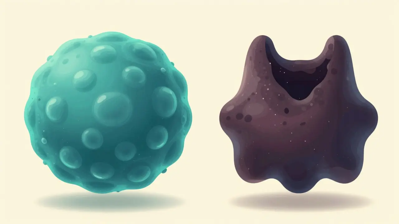
Benign nodules, by contrast, usually look round, have smooth edges, and often show a "spongiform" appearance, like a bubble wrap texture.
| Feature | Benign Indicators | Malignant Indicators |
|---|---|---|
| Echogenicity | Isoechoic or Hyperechoic (same or brighter) | Hypoechoic (darker) |
| Margins | Smooth, well-defined | Microlobulated, irregular, invasive |
| Composition | Cystic or Spongiform | Solid with microcalcifications |
| Growth | <2mm per year | >2mm per year |
| Lymph Nodes | Absent or Normal | Enlarged or Abnormal shape |
When Is a Biopsy Truly Needed?
This is the question patients ask most. Should I get stuck just to be sure? There is a reason why doctors hesitate to biopsy tiny spots. Over-diagnosis was a massive issue in the last decade. Many tiny cancers were found that would never have hurt anyone, leading to unnecessary surgeries with permanent side effects.
The Size Thresholds
We follow guidelines established by the American Thyroid Association, updated recently to reflect better outcomes. The decision tree relies on the ultrasound image score combined with nodule size:
- If a nodule looks suspicious, a biopsy is needed if it reaches 1 cm.
- If it looks moderately suspicious, wait until it hits 1.5 cm.
- If it looks benign, even a 2 cm nodule usually doesn't need a stick test unless you have a strong family history.
We also consider location. Nodules deep in the back of the neck near vital nerves might be watched longer to avoid complications. Also, remember that cysts larger than 3 cm causing compression symptoms often get treated, not just sampled.
The Biopsy Procedure: Fine-Needle Aspiration
Fine-Needle Aspiration (FNA)A minimally invasive procedure using a thin needle to extract cells from a thyroid nodule for analysis.
When the time comes, you need to know what happens in the room. This is called Fine-Needle Aspiration. It sounds scarier than it feels. The doctor uses ultrasound guidance to see the needle tip on a screen while guiding it directly into the lump. You might feel a pressure pinch, but the needle is very thin, similar to a blood draw.
Accuracy depends on skill. Learning curve analysis shows that it takes a physician about 50 to 75 supervised cases to hit a 90% adequacy rate. If you go to a community clinic, ask how often their doctors perform this specific procedure. If the sample isn't good enough (nondiagnostic), about 15 to 30% of the time, you'll simply repeat the procedure rather than rushing to surgery.

Decoding the Bethesda System Results
Once your cells are analyzed, you receive a report using the Bethesda System for Reporting Thyroid Cytopathology. It assigns your result to one of six categories. Knowing these risks helps you manage anxiety before talking to your surgeon.
| Category | Name | Risk of Cancer | Management Recommendation |
|---|---|---|---|
| Bethesda I | Nondiagnostic | 1% - 4% | Repeat FNA |
| Bethesda II | Benign | 0% - 3% | Clinical Monitoring |
| Bethesda III | Atypia of Undetermined Significance | 5% - 15% | Molecular Test or Repeat FNA |
| Bethesda IV | Follicular Neoplasm | 15% - 30% | Molecular Test or Diagnostic Surgery |
| Bethesda V | Suspicious for Malignancy | 60% - 75% | Diagnostic Thyroidectomy |
| Bethesda VI | Malignant | 97% - 99% | Total Thyroidectomy |
The grey areas are Category 3 and 4. Historically, these "indeterminate" results led to diagnostic lobectomies just to be safe, which means cutting out half your thyroid. Up to 35% of those surgeries revealed the lump was actually benign. This changed the game in recent years thanks to genetic testing.
Molecular Testing: Beyond the Basics
If your biopsy falls into that middle ground, your doctor might recommend a genomic classifier. Tests like Afirma GSC or ThyroSeq v3 analyze the DNA inside the nodule cells. They look for specific mutations known to cause thyroid cancer. If the test is negative, you can safely avoid surgery in 97% of cases. This technology has reduced unnecessary thyroidectomies by roughly 30% in major health centers.
While effective, these tests cost money. Medicare reimbursement for molecular testing ranges from $3,000 to $4,500 depending on the panel. Insurance approval varies, so financial counseling is worth discussing upfront. Despite the cost, avoiding a lifetime of thyroid hormone replacement is a strong trade-off for many.
Living with a Diagnosis
If everything checks out as benign, does it need removing? Often, the answer is no. Unless the nodule compresses your windpipe or esophagus, watchful waiting is standard. You might have an ultrasound checkup once a year or two. For those who do have confirmed cancer, treatment is highly effective. Papillary carcinoma, which makes up 80% of cases, often grows slowly and has excellent survival rates. New data from 2021 suggests active surveillance for cancers smaller than 1 cm is safe, with 87% remaining stable over five years.
Is pain a reliable sign of thyroid cancer?
Generally, thyroid cancer does not cause pain until it is very advanced. Painful nodules are more often associated with bleeding inside a benign cyst or thyroiditis (inflammation). However, rapid growth accompanied by pain should always be evaluated by a specialist.
How accurate is the ultrasound scan?
High-quality ultrasound is very sensitive. Studies indicate a sensitivity of 87% and specificity of 85% for detecting malignant nodules. Accuracy depends on the radiologist’s experience in reading thyroid images specifically.
Can a thyroid nodule shrink on its own?
Some nodules, particularly cystic ones, can decrease in size spontaneously. Radiofrequency ablation is also an option for shrinking symptomatic benign nodules, achieving volume reduction up to 78% at one year in recent trials.
Do I need to stop taking iodine or supplements?
You generally do not need to stop unless specifically advised. Excessive iodine intake can trigger dysfunction, but typical dietary amounts are safe. Always consult your endocrinologist before starting any new thyroid-specific supplements.
What happens if my initial biopsy is “nondiagnostic”?
This occurs in about 15% to 30% of biopsies due to lack of adequate cells. The standard protocol is to repeat the ultrasound-guided aspiration, ideally at a center with high procedural volume to improve the success rate.


Hope Azzaratta-Rubyhawk
April 1, 2026 AT 20:21We MUST embrace the facts presented here! It is crucial to understand that most nodules are harmless. Many people panic unnecessarily when a lump is found. Statistics clearly show that the majority remain benign throughout life. We should trust the medical guidelines provided in this post. Ignorance often leads to greater fear than the condition itself. Taking action like biopsies is necessary when thresholds are met. There is no reason to suffer in silence regarding your neck health. Modern technology provides us with amazing diagnostic tools. Ultrasound features help distinguish danger from safety effectively. Growth rates are significant indicators that doctors monitor closely. We celebrate the advancements in molecular testing for indeterminate cases. Avoiding unnecessary surgery protects patients from permanent side effects. Living with a diagnosis does not mean you cannot thrive afterward. Remember that surveillance is a valid path for small stable cancers.
Rob Newton
April 2, 2026 AT 05:04Most of this medical advice is completely useless garbage.
The Charlotte Moms Blog
April 2, 2026 AT 22:03I strongly disagree with your lack of insight!!! You are ignoring the clear benefits!!! Why would anyone dismiss the science??? It is reckless behavior!!!
Aysha Hind
April 3, 2026 AT 10:47The mainstream narrative hides deeper truths about thyroid health.
Hudson Nascimento Santos
April 4, 2026 AT 17:12Perhaps the truth lies in the gray areas we observe.
Dipankar Das
April 5, 2026 AT 21:18It is vital to maintain confidence in medical protocols.
sophia alex
April 6, 2026 AT 19:13They want us scared so we pay more!! 😱😱😱 Healthcare is failing us daily!!!
Mark Zhang
April 7, 2026 AT 02:45I hope everyone finds peace with their diagnoses.
simran kaur
April 7, 2026 AT 18:14Peace is a lie told by the pharmaceutical companies to keep us quiet.
Jenna Carpenter
April 8, 2026 AT 08:29This is junk infomation bout thyroids n biopsys.
Brian Shiroma
April 9, 2026 AT 07:41You really believe that because you read a headline somewhere?
Rachelle Z
April 9, 2026 AT 15:03Haha so funny!!! Everyone needs to relax!!!! 🙄🙄🙄 Seriously tho read the paper!!!
Branden Prunica
April 10, 2026 AT 09:12The world is ending and we talk about thyroid nodes!!
Divine Manna
April 11, 2026 AT 21:56The discussion regarding diagnostic thresholds requires rigorous examination. One must consider the nuance of cytological classification. It is undeniable that risk stratification saves lives. We cannot ignore the statistical significance of microcalcifications. The literature supports the current approach to management. A deviation from standard care could lead to poor outcomes. Therefore, adherence to established guidelines is paramount. Patients must understand the rationale behind monitoring protocols. Education empowers individuals to participate in their care plan actively. Without this knowledge, anxiety fills the void left by uncertainty. Trust in the medical process fosters better recovery metrics. Delaying necessary intervention can compromise long-term prognosis. Conversely, overtreatment introduces avoidable morbidity for the patient. Balance remains the central theme in modern endocrinology practice. We proceed with caution guided by empirical evidence.