Retinal Detachment: Emergency Symptoms and Surgical Treatment You Can't Afford to Ignore

When you notice a sudden shower of floaters, or a flash of light in your peripheral vision, it’s easy to brush it off as eye strain or aging. But if you’re experiencing these symptoms alongside a dark shadow creeping across your sight, retinal detachment could be happening - and every minute counts.
Retinal detachment isn’t a slow, gradual problem. It’s a medical emergency that can steal your vision permanently if not treated within hours. The retina, a thin layer of light-sensitive cells at the back of your eye, detaches from its blood supply. Without that connection, photoreceptors start dying. Once they’re gone, they don’t come back. That’s why speed isn’t just important - it’s everything.
What Are the Real Warning Signs?
You don’t need to be an ophthalmologist to recognize the red flags. Six symptoms appear suddenly and together - not one at a time.
- Sudden increase in floaters: Not just one or two. Patients describe seeing dozens of new dark spots, squiggles, or cobwebs that appear out of nowhere. The National Eye Institute says this isn’t normal aging - it’s a sign the vitreous gel inside your eye is pulling on the retina.
- Flashes of light: These aren’t the kind you get when you rub your eyes. They’re brief, bright streaks, often in your side vision, like a camera flash going off in the corner of your eye. They happen because the retina is being tugged.
- A dark curtain or shadow: This is the most urgent sign. It starts in your peripheral vision and slowly moves inward, like a shade being pulled across your field of view. If it reaches the center, your central vision is at risk.
- Blurry or distorted vision: Things look warped, wavy, or out of focus - even if you’re wearing glasses. This often means the macula, the part of the retina responsible for sharp vision, is already detached.
- Loss of side vision: You might bump into things on your left or right without realizing it. This isn’t just poor eyesight - it’s your retina failing.
- Sudden color changes: Colors look washed out, especially if the macula is involved. It’s not a lighting issue - it’s your retina losing function.
These symptoms don’t come and go. They stick. And they worsen. A 2022 study in the Journal of VitreoRetinal Diseases found that patients treated within 24 hours had a 90% chance of successful reattachment. Wait 72 hours, and your chance of keeping 20/40 vision drops from 75% to 35%.
How Is It Diagnosed?
There’s no home test. No app. No over-the-counter remedy. Diagnosis requires specialized tools and expertise.
An eye doctor will dilate your pupils and use an indirect ophthalmoscope - a bright light with a special lens - to look directly at the retina. If the view is blocked by blood or cloudiness, they’ll use B-scan ultrasound to create a real-time image of the back of your eye. Optical coherence tomography (OCT) gives a high-res cross-section of the retina, showing exactly where and how deep the detachment is.
These aren’t standard tools. Most family doctors or urgent care clinics don’t have them. That’s why 63% of patients in one American Society of Cataract and Refractive Surgery survey were misdiagnosed with “eye strain” before being referred to a specialist. If you have these symptoms, go straight to an eye doctor who specializes in retinal conditions - not your general practitioner.
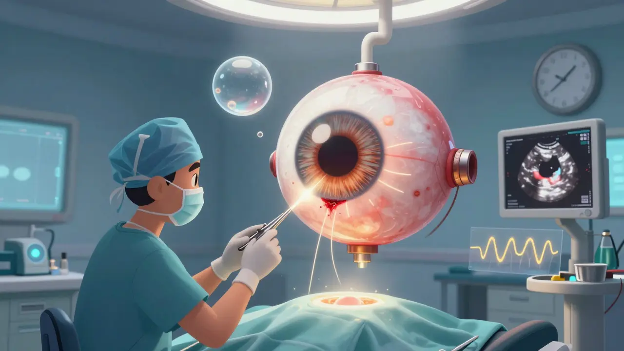
What Are the Surgical Options?
There are three main surgeries, each with pros and cons. The choice depends on the size, location, and complexity of the detachment.
Pneumatic Retinopexy
This is the least invasive. A gas bubble is injected into the eye. You then position your head so the bubble floats up and presses against the detached area, sealing the tear. Laser or freezing treatment is used to weld the retina back in place.
It works well for small, single tears near the top of the retina - about 70-80% success rate. But it won’t work if the tear is below the center of the eye. And you’ll need to keep your head in a specific position - often face-down - for 50% of every day for up to 10 days. That’s hard to do, especially if you work, drive, or have young kids.
Scleral Buckling
This involves sewing a soft silicone band around the outside of your eye. It gently pushes the wall of the eye inward, helping the retina reattach. It’s often used for younger patients or those with lattice degeneration - a thinning of the retina.
Success rates are high: 85-90%. But it can cause side effects. About 1.5 to 2.0 diopters of nearsightedness are common afterward. Some people develop double vision. And it doesn’t work well if the detachment is large or if scar tissue has already formed.
Vitrectomy
This is the most common surgery today - used in 65% of cases, according to the American Society of Retina Specialists. The surgeon removes the vitreous gel from inside your eye and replaces it with a gas bubble or silicone oil. The bubble presses the retina back into place, and laser or freezing seals the tear.
Vitrectomy has the highest success rate - 90-95% - especially for complex cases, giant tears, or when the macula is involved. But there’s a big trade-off: if you still have your natural lens, you’ll likely develop a cataract within two years. About 70% of patients do. You’ll need cataract surgery later.
There’s also a newer version - minimally invasive 27-gauge vitrectomy - approved in early 2023. It uses smaller incisions, less trauma, and faster healing. But it’s only available at major eye centers.
Why Timing Is Everything
Dr. Carl Regillo, Chief of Retina at Wills Eye Hospital, says: “Every hour counts.”
When the macula detaches - the part of the retina you use to read, drive, and recognize faces - vision loss is rapid. Studies show visual recovery drops by about 5% per hour after symptoms start. If you wait 48 hours, your chance of seeing 20/40 or better is cut in half.
Wills Eye’s emergency protocol requires patients with macula-off detachments to be seen within 4 hours. Surgery must happen within 12. That’s not a suggestion - it’s the standard of care.
One Reddit user, "VisionWarrior22," ignored floaters for three days. By the time they got help, the shadow had reached the center of their vision. Their final acuity was 20/100 - far worse than the 20/25 they could’ve had with same-day treatment.
What Happens After Surgery?
Recovery isn’t quick. And it’s not comfortable.
If you had a gas bubble, you’ll need to keep your head in a specific position - often face-down - for 7 to 10 days. You can’t lie on your back. You can’t drive. You can’t read normally. Many patients need help with meals, hygiene, and childcare. A 2022 survey found 41% of patients reported significant discomfort from positioning.
Post-op complications include:
- Cataracts (70% within two years after vitrectomy)
- High eye pressure (25% of cases)
- Re-detachment (5-15%, depending on technique)
- Infection (less than 1%, but serious)
Follow-up visits are frequent. You’ll need checkups at 1 day, 1 week, 1 month, and then every few months for a year. Your eye pressure, retina position, and vision will be tracked closely.
Who’s at Risk?
Retinal detachment affects about 1 in 10,000 people each year. But some groups are far more vulnerable:
- People with severe nearsightedness (over -5.00D): 167 in 10,000 annual risk
- Those who’ve had cataract surgery: 0.5% to 2% risk
- People with lattice degeneration: 1% lifetime risk
- Those with a family history of retinal detachment
- People who’ve had eye trauma or previous retinal detachment in the other eye
Age matters too. Risk jumps after 40. But it can happen at any age - especially after injury or in people with genetic conditions.
What About Prevention?
There’s no guaranteed way to prevent retinal detachment. But you can reduce your risk:
- Get regular eye exams - especially if you’re over 40 or nearsighted
- Wear protective eyewear during sports or risky activities
- Know your family history
- Don’t ignore sudden floaters or flashes - even if they seem minor
Some doctors debate whether to treat asymptomatic lattice degeneration. A 2021 study suggested prophylactic laser could help high-risk patients. But others warn the procedure carries its own risks - and most people with lattice will never develop detachment.
The bottom line? Don’t wait for a doctor to tell you something’s wrong. If you notice sudden changes in your vision, act.
What If You Can’t Get to a Specialist Right Away?
If you’re in a rural area - and only 35% of U.S. counties have a retinal specialist - and you can’t get to a clinic within hours, go to the nearest emergency room. Ask for an ophthalmology consult. Bring up retinal detachment by name. Say: “I have symptoms of retinal detachment and need immediate evaluation.”
Many hospitals can do basic exams with portable ultrasound. Even if they can’t do surgery, they can stabilize you and arrange transfer to a center that can.
Don’t wait for a referral. Don’t schedule an appointment for next week. This isn’t a cold or a headache. It’s an emergency that can end your vision forever.
What’s Next for Treatment?
Technology is improving. Intraoperative OCT - real-time imaging during surgery - is now helping surgeons remove scar tissue more precisely. AI tools are being tested to spot early signs of detachment in routine eye scans, potentially catching cases before symptoms appear.
Long-term, researchers are working on bioengineered retinal patches and gene therapies for inherited conditions that predispose people to detachment. But those are still years away.
For now, the best tool we have is awareness. Know the symptoms. Act fast. Get to the right specialist. Your vision depends on it.
Can retinal detachment fix itself?
No. Retinal detachment cannot fix itself. The retina needs to be physically reattached by a surgeon. Without treatment, the photoreceptor cells die permanently, leading to irreversible vision loss. Even if symptoms seem to improve, the detachment is still there and will worsen.
Is retinal detachment surgery painful?
The surgery itself is not painful - it’s done under local or general anesthesia. Afterward, you may feel pressure, mild discomfort, or a scratchy sensation. Most patients report manageable pain with over-the-counter medicine. The bigger challenge is the positioning requirement after surgery, especially if you need to stay face-down for days.
How long does recovery take?
Initial healing takes about 2 to 4 weeks, but full recovery can take months. Vision may remain blurry for weeks as the gas bubble dissipates. If you had a gas bubble, you can’t fly or go to high altitudes until it’s gone - which can take 6 to 8 weeks. Your final vision result depends on whether the macula was detached and how quickly you were treated.
Can I drive after retinal detachment surgery?
No - not until your doctor clears you. If you had a gas bubble, your vision will be distorted and your peripheral vision limited. You also can’t fly. Most patients can’t drive for at least 2 to 6 weeks, depending on the procedure and healing. Your doctor will give you specific guidelines based on your case.
Will I need glasses after surgery?
You may. Scleral buckling often causes new nearsightedness. Vitrectomy can speed up cataract development, which will require glasses or future surgery. Even if your vision returns, your prescription might change. Most patients need updated glasses or contacts after recovery.
What’s the success rate of retinal detachment surgery?
The anatomical success rate - meaning the retina is reattached - is 85-95% with modern techniques. But functional success - meaning good vision returns - depends on timing. If the macula was detached for less than 24 hours, most patients regain 20/40 vision or better. If it was detached for more than a week, vision may remain permanently impaired, even if the retina is successfully reattached.


Vince Nairn
January 6, 2026 AT 14:36Also, if you're in a small town, just go to ER and say 'retinal detachment' like it's a code word. They'll scramble.
Aparna karwande
January 8, 2026 AT 13:50Jessie Ann Lambrecht
January 9, 2026 AT 13:59Pro tip: buy a face-down pillow. It's a game-changer.
Ayodeji Williams
January 10, 2026 AT 09:25got surgery 3 days later and now i see 20/50 instead of 20/200. still better than blind tho 🙏
Elen Pihlap
January 11, 2026 AT 04:16Paul Mason
January 11, 2026 AT 16:58Katrina Morris
January 13, 2026 AT 01:05steve rumsford
January 13, 2026 AT 06:45LALITA KUDIYA
January 13, 2026 AT 11:51Anthony Capunong
January 13, 2026 AT 23:44Kyle King
January 15, 2026 AT 21:23Poppy Newman
January 17, 2026 AT 07:25Sai Ganesh
January 18, 2026 AT 03:56